

Alexandre Simões Barbosa1,2
DOI: 10.17545/eOftalmo/2026.v12.0017
ABSTRACT
The low vision clinic entails the integrated assessment of the functional aspects of the visual experience, especially visual acuity as high-contrast discrimination, on-axis and off-axis refraction, the identification of low- and high-order aberrations, the point spread function, the contrast sensitivity curve, the location and stability of the preferred retinal locus, the assessment of glare and crowding, and reading parameters. This characterization across different domains allows for a consistent functional analysis that forms the basis for the rehabilitation strategies in each patient, given the demands for efficiency and autonomy. This chapter aims to discuss the fundamental elements for a structured clinical assessment that allows for the management of low vision rehabilitation, with special emphasis on patients with afferent and integration pathway lesions. Due to the complexity of the visual experience, rehabilitation strategies aim to treat the retinal image, stabilize the fixation, and improve efferent oculomotor control, enabling greater efficacy of presaccadic programming and scanning movements. Faced with the challenge of rehabilitation, especially in neuro-ophthalmology, this article proposes a structured approach centered on the refinement of the functional aspects of the visual experience.
Keywords: Low vision; Visual rehabilitation; Preferred retinal locus; Fixation stability; Contrast sensitivity
RESUMO
A clínica da baixa visão pressupõe a avaliação integrada dos aspectos funcionais da experiência visual, sobretudo a acuidade visual como discriminação em alto contraste, a refração on-axis e off-axis, a identificação de aberrações de baixa e alta ordens, o point spread function, a curva de sensibilidade ao contraste, a localização e estabilidade do locus retiniano preferencial, a pesquisa do glare e do crowding, e os parâmetros de leitura. Essa caracterização em diferentes domínios permite uma análise funcional consistente que constitui a base para as estratégias de reabilitação em cada paciente, diante das exigências de eficiência e autonomia. Esse capítulo tem por objetivo discutir os elementos fundamentais para uma avaliação clínica estruturada que permita o manejo da reabilitação em baixa visão, com especial ênfase em pacientes com lesão de vias aferentes e de integração. Devido à complexidade da experiência visual, as estratégias de reabilitação visam não apenas o tratamento da imagem retiniana, como também a estabilização da fixação e aprimoramento do ajuste eferente, possibilitando uma maior eficácia da programação pré-sacádica e movimentos de varredura. Diante do desafio da reabilitação, especialmente em neuro-oftalmologia, o capítulo propõe uma abordagem estruturada centrada no refinamento dos aspectos funcionais da experiência visual.
Palavras-chave: Baixa visão; Reabilitação visual; Locus retiniano preferencial; Estabilidade de fixação; Sensibilidade ao contraste
INTRODUCTION
The characterization of the different patterns of functional loss in each patient with low vision requires a structured assessment. Low vision rehabilitation practice is based on a wide variability of functional impairments, often multiple, that present when faced with demands for independent living. In low vision practice, and especially in neuro-ophthalmology rehabilitation, the particularities of each case allow us to revisit the applicability of current intervention models, tailoring rehabilitation modalities to the specific needs of each patient. In this context, it is not sufficient for the person with low vision to be seen by a multidisciplinary team. Moreover, the ophthalmologist or neuro-ophthalmologist who delves into this field needs a comprehensive knowledge of the visual phenomenon, including the determinants of the retinal image, the establishment of foveal and extrafoveal fixation areas, the dynamics of saccadic fixation and refixation movements, and the integration of afferent visual inputs, in order to conduct an effective rehabilitation process. This article aims to address these fundamental points of neuro-ophthalmology rehabilitation.
STRUCTURED EVALUATION OF VISUAL FUNCTIONS
Evaluation of the optic pathways
Medical History
The history of low vision must be carefully revisited for a robust review of the diagnosis, the progression of low vision, effects of medical interventions, and sequential analysis of supplementary tests1,2. The history must include fundamental autobiographical elements, such as education, professional and intellectual activity, level of engagement with work, and significant professional and personal ties, projects, and expectations1,2. Previous rehabilitation experiences, their challenges, and outcomes are fundamental; so, these experiences can be incorporated into new strategies.
Visual acuity
A structured examination of visual functions begins with the visual acuity obtained with best correction, which is defined as the function of discriminating two distinct points in high contrast and constitutes a fundamental parameter in the strategy for optical aid fitting. Improvements considered small, less than a one-line gain on the ETDRS chart, make a relevant difference in rehabilitation strategies2. Retinoscopy is indispensable for an adequate refraction and can suggest higher-order aberrations2. The refraction should be finalized with a trial frame2. If the patient has extrafoveal fixation, the refraction should be performed with this fixation, which introduces fundamental paraxial elements for an adequate optical correction3. A careful refraction improves the quality of the retinal image for high- and low-contrast optotypes, producing a significant overall gain in visual function1-7.
Contrast sensitivity function
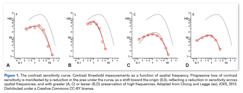
The contrast sensitivity curve constitutes the graphical representation of the contrast threshold as a function of spatial frequency, measured in cycles per degree, corresponding to the widths of alternating black and white bands2,7-9. The area under the contrast sensitivity curve corresponds to the integral of the contrast sensitivity function with respect to spatial frequency and constitutes a robust parameter of visual function, even in patients with preserved visual acuity at high contrast1-4,7,8. Different area measurements can occur despite 20/20 visual acuity. Areas of different contrast sensitivities can be identified within scotomas, especially central ones, enabling fixation2,6,10,11. From the fovea, there is an exponential decay of contrast sensitivity, which hinders stable fixation3,12,13. Accordingly, isopters can be understood as spatial representations of regions delimited by the same contrast threshold. The contrast sensitivity curve is of particular importance in neuro-ophthalmology, capable of identifying afferent lesions compatible with the preservation of visual acuity at high contrast1,2,7,8. Examples of contrast sensitivity curves are shown in figure 1.
The preferred retinal locus
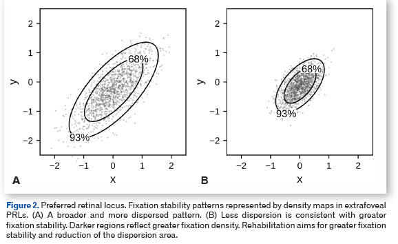
Foveal or papillomacular bundle damage implies the development of an extrafoveal preferred retinal locus (PRL). The quality of the image obtained from the PRL is determined by its location, extent, and contrast sensitivity2,11,14,15. The PRL can be located inside or outside central scotomas, and this distinction will be fundamental in the choice of rehabilitation strategies2,10,11,16,17. The characterization of the PRL is done through microperimetry, but its presence can be suggested by automated perimetry, where the central scotoma appears displaced on the visual field plot, along with the blind spot2,11,12. Although the use of the PRL and its location are unconscious, training techniques with tangent screens can be used for its recognition and improvement2,9,14. Graphic representations of the PRL with different densities and dispersion areas are shown in figure 2.
Visual fields
The location and extent of visual field defects are the main determinants of both the limitations imposed by afferent lesions and the proposals for their rehabilitation. A wide variability of field defects can be classified into central and peripheral. However, central defect may be associated with islands of central fixation, while peripheral defect may extend to paracentral regions. In addition to the location and extent of the scotoma, contrast sensitivity at its margins is fundamental in the response to rehabilitation strategies1,2,10. Furthermore, the location of the scotoma margins depends on luminance, and low luminance manifests as an expansion of the scotoma area1,2,6.
Islands of greater preservation of retinal sensitivity allow the development of PRLs within the scotoma, as in Leber hereditary optic neuropathy (LHON)2,10,16. Automated perimetry methods, which assume stable foveal fixation, may present artifacts produced by fixation from an extrafoveal PRL, especially in the presence of unstable fixation2,12. Furthermore, central scotomas, due to their extensive cortical representation, cause significant limitation in orientation and mobility.
An eccentric location of the central scotoma in 30-2 strategies, associated with a displacement of the blind spot, is compatible with an extrafoveal PRL. In these cases, supplementation with 10-2 strategies should be considered. However, the method of choice for the evaluation of central scotomas is microperimetry, because it allows the stimulus to be projected without the interference of microsaccadic movements, corrected through eye-tracking2,6,10,18. Microperimetry allows the direct identification of the PRL and the evaluation of fixation stability, contributing to better understanding of reading dynamics2,11,17-19.
Hemifield defects often present with bitemporal or homonymous patterns, due to chiasmal and postchiasmal lesions. These defects do not involve the central field and are therefore considered peripheral. However, paracentral or central defects may occur, which changes the presentation of these lesions. Chiasmal lesions can produce paracentral bitemporal defects, while occipital pole lesions can produce homonymous defects restricted to the central hemifield. Complete hemifield defects and hemifield defects with macular sparing show significant differences regarding limitations for reading activities1,12,20. Ideally, Goldmann perimetry associated with computerized visual field testing or microperimetry in selected cases should be used for the characterization of field defects 2,6,10.
Reading parameters
Reading allows the assessment of multiple visual functions together, in addition to attention and cognitive functions, which are fundamental for rehabilitation. During reading, scanning the text demands fixation and refixation movements that reflect the structural status of the fovea or the PRL, especially contrast sensitivity and the extent of the area available for fixation11,14,19.
Fluent reading requires a foveal area of approximately 7º of visual angle, asymmetrically distributed, with 5º in the direction of reading and 2º in the opposite direction12,13. Reading performance requires the simultaneous integration of foveal and parafoveal information, allowing for the anticipation and planning of the subsequent saccadic movement11,12,20. Reading also requires some suppression of the crowding effect, whereby the presence of nearby objects, defined by line, word, and letter spacing, hinders the perception of margins and segregation of images19,21.
The crowding effect can be assessed by comparing the visual acuity obtained with isolated optotypes to that obtained with optotypes presented together, close to each other, in space. The loss of contrast of the retinal image contributes to the crowding effect by reducing the definition of its margins.
The evaluation of reading quality should be based on its speed, word substitution rate, and fluency1,2,20. Reading speed constitutes a global parameter that involves saccadic programming, fixation stability, and the interposition of scanning (return-sweep) movements and regressive movements for fixation2,19,20. Presaccadic programming constitutes the process of anticipating the next refixation movement, in which paracentral information modulates the direction and amplitude of the saccadic movement, producing effective navigation through the text. The behavior of reading parameters under high- and low- contrast conditions adds information about subclinical losses of image stabilization and saccadic programming.
Optical coherence tomography
Functional and structural parameters should be compared in lesions of the optic pathways, especially the contrast sensitivity curve and visual fields with measurements of the peripapillary retinal nerve fiber layer (pRNFL) and macular ganglion cell-inner plexiform layers (GC-IPL). Structural loss may not be identified in functional tests alone, such as automated visual field testing (standard automated perimetry) and Goldmann perimetry, but they compromise performance in more complex functional tests, such as the contrast sensitivity curve or low-contrast reading1,2,6-8. However, the correlation between functional measurements and arameters may be inconsistent2,6,10,18.
Patients with anti-MOG antibody-associated optic neuritis (MOGAD) present with relative preservation of the visual field and contrast sensitivity that are not suggested by the significant reduction in GC-IPL thickness. On the other hand, patients with Leber hereditary optic neuropathy associated with 14484 (ND6 mitochondrial DNA) and JC30 (autosomal recessive) mutations may present with significant reduction in the scotoma density despite progression of structural loss. Therefore, a refined structural assessment of the optic nerve does not replace a functional assessment, in which the thresholds for response can be adjusted2,6,22. For all afferent lesions, pRNFL and GC-IPL measurements of less than 50 and 35 µm are less consistent, due to the floor effect. These findings demonstrated the fundamental importance of the joint assessment of structural and functional parameters2,6,10,16.
DETERMINANTS OF THE RETINAL IMAGE
Corneal topography and wavefront analysis
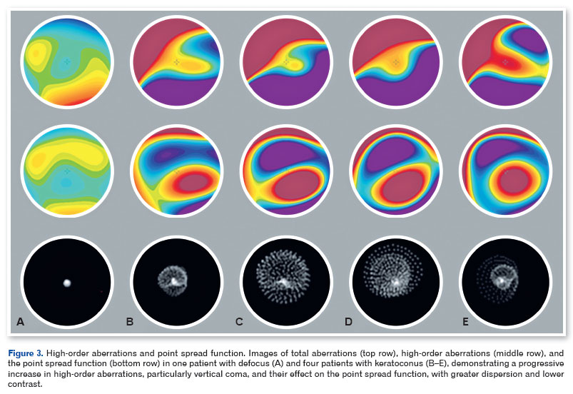
Corneal topography and wavefront analysis are fundamental elements in the evaluation of patients with low vision and evidence of high-order aberrations, especially in those with extrafoveal fixation. By wavefront analysis, the contribution of low-order (defocus and astigmatism) and high-order (spherical aberration, high-order astigmatism, and coma) aberrations to retinal image formation can be evaluated2,4,5. Special emphasis should be given to high-order aberrations, which can be produced by the geometric decentration associated with the PRL even in emmetropic patients, and which are not correctable with glasses2,3,5. High-order aberrations manifest as light scatter, especially with the increase in pupillary diameter4,5. Patients with extrafoveal fixation should assume this fixation during the acquisition of topographic images for proper evaluation of the off-axis map of aberrations2,3,5. While on-axis refraction is oriented by the visual axis established by foveal fixation, off-axis refraction is oriented by the PRL and expresses the refractive effects and high-order aberrations determined by this change of axis. Total and high-order aberration maps in myopia and in distinct stages of keratoconus are shown in figure 3, along with their respective point spread function (PSF) diagrams.
Glare
Glare can be defined as the reduction in the contrast of the retinal image caused by the intraocular light scatter, producing a broadening of the PSF and a degradation of the contrast sensitivity function, especially intermediate and high frequencies1,4,5,23. This effect is wavelength λ-dependent and occurs primarily at λ values below 450 nm1,2,5. Selective (yellow) filters act on λ <450 nm (blue) and reduce glare, producing an increase in contrast of the retinal image1,2,5,17. Distinct filters, such as in the λ ~ 511 nm (amber) and λ ~ 525 nm (orange) range, can be used for more intense glare in high-luminance situations. In any case, the clinical test based on the comparison of the contrast sensitivity curve with different filters should be used as the selection criterion for specific luminance conditions. Some patients show significant improvement in contrast sensitivity with filters even in the absence of glare-related complaints.
Point spread function
A PSF provides a direct representation of retinal image quality from a map of the light energy distribution produced by the effect of scattering from a point source of light4,5,23,24. Parte superior do formulário
Parte inferior do formulário
Refraction and correction of higher-order aberrations aim to enhance the retinal image, reducing this scattering. Image processing in rehabilitation aims to obtain a smaller scattering pattern, producing an image with higher luminous density and greater contrast4,5,23,24. An inconsistent or paradoxical response to optical aids may result from greater scattering, especially when irregular, from the point of incidence. Thus, rehabilitation proposals must be considered based on the analysis of the PSF and the refinement of retinal image. The point-spread diagrams associated with defocus and higher-order aberrations are shown in figure 3.
REHABILITATION STRATEGIES
Optical aids for near vision
The best corrected visual acuity should be the starting point for choosing near optical aids. According to Kestenbaum's rule, the reciprocal of the distance best-corrected visual acuity provides an estimate, in diopters, of the plus add required for fluent reading at the standard near working distance (e.g., ~25 cm), corresponding to 1M print2. However, this rule underestimates magnification when there is significant loss in contrast sensitivity, and it assumes foveal fixation, without the restrictions imposed by the establishment of a PRL2,11,12. The analysis of reading quality must be performed with different magnifications, starting from the one proposed by Kestenbaum's rule, aiming for the lowest magnification capable of producing fluent reading. Beyond this point, additional magnification compromises fluency, due to the limitation of the field and saccadic programming. Non-optical resources should be tried, especially in patients with low contrast sensitivity, such as selective filters or typoscopic cards, in addition to lighting adjustment1,9,13,17. The reading test identifies patients who are candidates for electronic aids, which offer higher magnification levels, without focal distance adjustment, in addition to allowing for brightness and contrast adjustments1,2,9,17.
Telesystems
Telesystems allow for magnification with focal distance adjustment as needed. Their main parameters, which define the quality of the image they produce, are the magnification power and the exit pupil diameter, indicated on the body of the telesystem. The magnification should be the lowest possible for a given activity to allow for greater field preservation and more effective scanning movements, especially in patients with peripheral field loss1,9,12,13. On the other hand, the exit pupil should be as large as possible up to the patient's pupil diameter, especially when there is a significant reduction in contrast sensitivity2,17,19,25.
Telescopic systems can accentuate the contribution of higher-order aberrations in retinal image formation, especially in the presence of extrafoveal PRL, by introducing off-axis2,3,5 components. Accordingly, in the presence of higher-order aberrations, the use of the telescopic system with and without correction with rigid contact lenses2,4,5,23 should be considered. The comparison of contrast curves in both situations allows for an objective assessment of the indication for these lenses. Furthermore, modifications in pupil diameter should be considered in the choice of telescopic systems, both due to the greater contribution of higher-order aberrations in larger pupillary diameters and for the adjustment of the exit pupil.
STRATEGIES BY FUNCTIONAL GROUPS
Central scotomas
The location of the field defect is the main determinant of rehabilitation strategies in patients with low vision. Among optic neuropathies, mitochondrial forms (such as hereditary and toxic-nutritional) and demyelinating forms (such as those associated with multiple sclerosis and MOGAD) produce central scotomas of varying densities and extents, and some of which exhibit islands of relatively preserved sensitivity that allow fixation through a new PRL6,16,17,20. While islands of vision within central scotomas present limitations to magnification, PRLs located at their margins generally show a favorable response to magnification2,12,13,17.
Adequate optical correction, especially that of higher-order aberrations, enhances the retinal image and increase in fixation stability5,10,11,23. The enhanced retinal image must then be subjected to magnification adjustment, starting from the positive lenses suggested by Kestenbaum's rule, down to the lowest magnification that allows for fluent reading. Reading parameters constitute the best criterion for magnification adjustment.
Rehabilitation includes training in awareness of the central scotoma and the PRL, as well as the saccadic movements necessary for fixation and refixation, by means of tangent screens or microperimetry, which increases the efficacy of saccadic movements, reading speed, and text fluency10,17,19,20. Especially in recent visual loss, the development of an inferior PRL, with elevation of the eye and superior displacement of the scotoma, constitutes the most favorable situation11,12,14,15.
In patients with central scotoma, the choice of telesystems should be based on their magnification and exit pupil parameters. Just as for near, the lowest magnification should be considered, comparing the contrast curve of different models with distinct magnification and exit pupil values2,17,19,25. The highest ratio between exit pupil and magnification should be chosen for the minimum magnification required for each activity. The effects of off-axis refraction correction and higher-order aberrations on the contrast sensitivity curve, as well as the effect of crowding of isolated and multiple optotypes, should be tested while choosing telesystems3-5,21,25.
Paracentral, arcuate, and ring defects
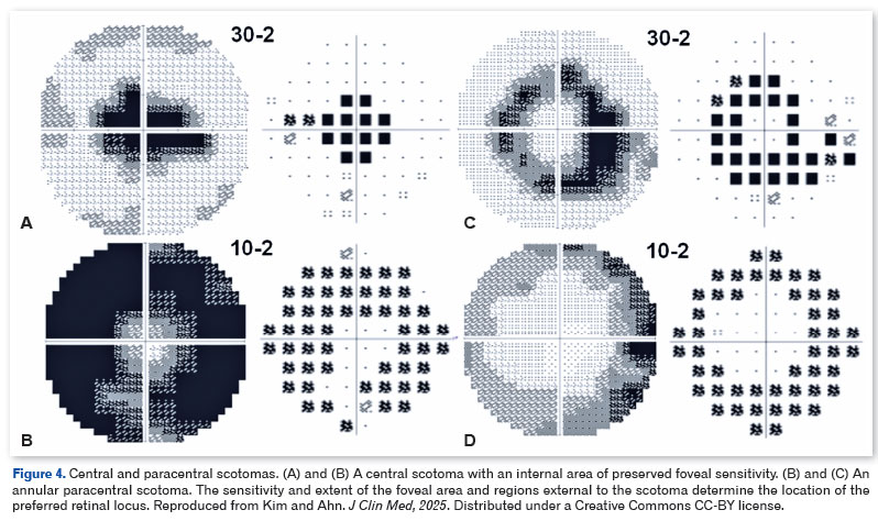
Paracentral and arcuate scotomas occur usually in ischemic and glaucomatous optic neuropathies and in inflammatory optic neuritis. In particular, ring scotomas can be produced by continuity from superior and inferior arcuate scotomas. Paracentral and arcuate scotomas impose significant restrictions on foveal and parafoveal integration, impairing saccadic programming1,12,13,20. The reduction in the area available for foveal fixation limits magnification and contributes to the establishment of a PRL external to the scotoma12,13,15. The possibility of both foveal and extrafoveal fixation, as well as of two distinct extrafoveal areas, determines the specialization of two PRLs for distinct activities14, which are established as a function of contrast sensitivity and the available extent for image projection2,11,14. Inferior arcuate scotomas impose greater restrictions on rehabilitation than superior ones, whereas in ring scotomas these restrictions are even greater by circumscribing the central area, separating the foveal fixation area from extrafoveal areas1,2,12,13,15. A central scotoma with an internal area of foveal sparing, an ring scotoma, and a peripheral field loss involving the mid-peripheral area are shown in Figure 4.
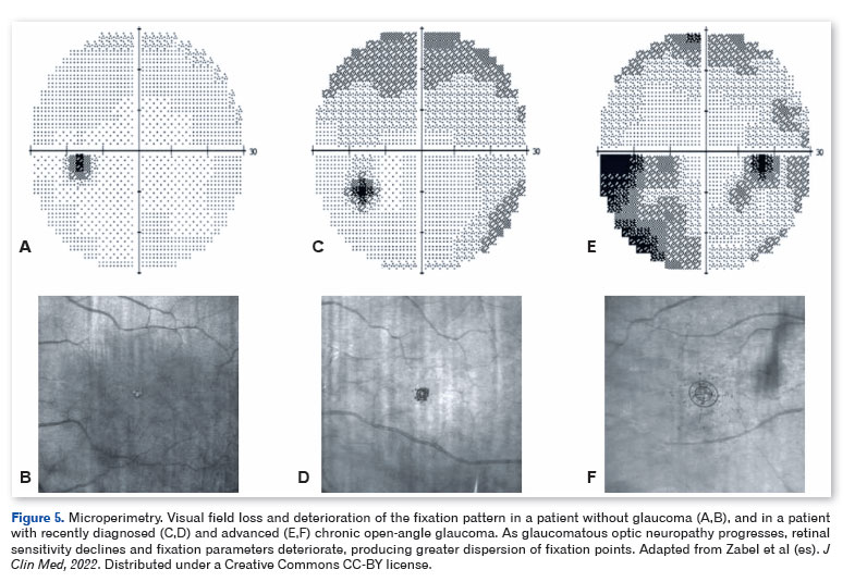
Due to the restrictions imposed by paracentral, arcuate, and an ring scotomas, strategies focused on retinal image enhancement and saccadic training yield better results than magnification1,12,13. Non-optical aids, such as reading guides and typoscopic cards, are often used for rehabilitation in arcuate scotomas, which promotes a more effective and stable fixation shift, as a preferred path between distinct fixation points1,2,13,14. In addition, ring scotomas can produce a paradoxical response to magnification, in which the enlarged retinal image overlaps the scotoma. The association between visual field losses and fixation instability in glaucoma is shown in figure 5.
Bitemporal, homonymous, and altitudinal visual field losses
Bitemporal and homonymous field defects constitute the signatures of chiasmal and retrochiasmal lesions, respectively. Posterior chiasmal lesions are especially important because they produce paracentral bitemporal field loss, while anterior or posterior extension of chiasmal lesions involve the nasal fields. Retrochiasmal lesions produce contralateral homonymous defects with preservation of the central field, while occipital pole lesions produce central homonymous defects. Altitudinal field defects are the most attributed to ischemic optic neuropathies, but they are also observed in other optic neuropathies, as well as lesions of the superior or inferior margin of the calcarine fissure.
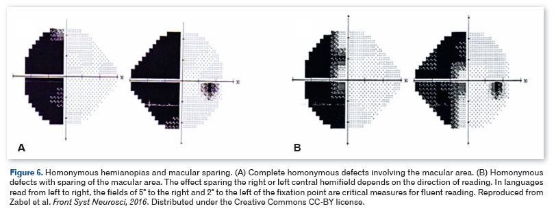
Extensive paracentral bitemporal defects can compromise reading due to their location to the right and left of the fixation point12,13. In addition, the absence of overlap of the temporal fields on convergence results in a lack of representation of the region posterior to the fixation point, with loss of fusion stability due to hemifield sliding11,12,17. As a rule, the presentation of hemianopsias depends not only on their extent and the involvement of the central and paracentral fields but also on their location in relation to the reading direction. In languages with left-to-right reading, right homonymous loss near the fixation point significantly interferes with reading progression, requiring a large number of scanning (return-sweep) and regressive movements, whereas left homonymous and inferior visual field defects impair line-finding12,13,19,20. Central homonymous scotomas related to lesions of the occipital pole produce a loss of half of the central field designated for reading to the right or to the left, with a significant limitation of reading12,13.
In central and paracentral homonymous field defects, a consistent compensatory strategy is the displacement of the fixation point toward the remaining field11,12,17,20. In a right homonymous field defect, the displacement of the fixation point to a PRL on the left implies an increase in the area available for reading. The improvement of the saccade pattern through training favors the replacement of unstable and dysmetric saccadic movements with more predictive and stable refixation movements12,14,20,21,26.
Orientation and mobility are particularly compromised in homonymous field loss, due to the suppression of saccadic movements of both the affected and the remaining hemifield, aggravated by different levels of neglect characterized by a lack of awareness of the visual field loss1,20,26. In patients with homonymous hemianopsia, saccadic training is the most effective form of rehabilitation for orientation and mobility, and is based on the execution of scanning saccadic movements toward the region of visual field loss, producing an improvement in the scanning pattern1,13,17,20,26.
Extensive peripheral defects, with preservation restricted to the central region, can be found in cortical lesions that spare the occipital pole. Two cases of homonymous losses, one with macular area involvement and another with macular area sparing, are shown in Figure 6. Rehabilitation follows the guidelines for the previously described ring scotomas, with the distinction that PRLs cannot be established outside the scotoma12,20. Occipital pole lesions behave like central scotomas, and their rehabilitation follows what was previously described1,13. The rehabilitation of cortical lesions aims, in addition to visual function, to reintegrate spatial perception, limiting the manifestations of neglect.
Disorders of visual integration
Balint's syndrome is the model of loss of spatial integration due to a parieto-occipital (dorsal) lesion responsible for object localization in space1,2,26,27. The syndrome is characterized by persistence of fixation, difficulty initiating voluntary saccades, and an inability to simultaneously represent more than one object in a scene2,20,26,27. The condition may manifest in patients with neurodegenerative conditions such as Alzheimer's disease1,27 and posterior cortical atrophy20,26,27. The clinical manifestations are disabling, and reading is severely impaired due to fragmentation of the text and saccadic inhibition1,20,26,27. Rehabilitation aims to improve saccadic exploration and scanning1,20.
Charles Bonnet syndrome is characterized by complex visual hallucinations associated with visual loss that are not attributable to coexisting neurological or psychiatric conditions13,29,30. The hallucinations are produced by hyperexcitability and spontaneous activity of the visual association areas29,30. Although no interventions are capable of suppressing the hallucinations, patient counseling and rehabilitation represent the most effective management 2,13,29,30. With rehabilitation, more afferent input replaces the silence of the integrative pathways, increasing the threshold for spontaneous activity that produces unreal images.
Closing remarks
Visual rehabilitation in neuro-ophthalmology is based on the structured assessment of visual functions, considering all determinants of the visual experience. Special attention must be given to the retinal image, which can be enhanced to allow for stable fixation. Optical and electronic aids must be individually tested based on a complete functional diagnosis, according to the demands of each case. The adaptation of these aids must be accompanied by saccadic eye movement training based on the recognition of the location of visual field loss and development of scanning strategies. Rehabilitation must be based on the specific knowledge developed for each case, which guides the decisions made as the rehabilitation progresses.
REFERENCES
1. Kerkhoff G. Restorative and compensatory therapy approaches in cerebral blindness: a review. Restor Neurol Neurosci. 1999; 15(2-3):255-71.
2. Markowitz SN. Principles of modern low vision rehabilitation. Can J Ophthalmol. 2006;41(3):289-312.
3. Navarro R, Losada MA. Aberrations and relative efficiency of ray pencils in the living human eye. Optom Vis Sci. 1997;74(7):540-7.
4. Guirao A, Porter J, Williams DR, Cox IG. Calculated impact of higher-order monochromatic aberrations on retinal image quality in a population of human eyes. J Opt Soc Am A Opt Image Sci Vis. 2002;19(1):1-9.
5. Lombardo M, Lombardo G. Wavefront aberration of human eyes and new descriptors of image optical quality and visual performance. J Cataract Refract Surg. 2010;36(2):313-331.
6. Kim KE, Ahn SJ. Visual field examinations for retinal diseases: a narrative review. J Clin Med. 2025;14(15):5266.
7. Chung ST, Legge GE. Comparing the shape of contrast sensitivity functions for normal and low vision. Invest Ophthalmol Vis Sci. 2016;57(1):198-207.
8. Richman J, Spaeth GL, Wirostko B. Contrast sensitivity basics and a review of currently available tests. J Cataract Refract Surg. 2013;39(7):1101-6.
9. Sánchez JM. Más allá de la baja vision. Rev Salud Bosque. 2017;1(2):49-58.
10. Kiszka A, Nowakowska D, Nowomiejska K, Tońska K, Rejdak R. Visual rehabilitation training in a patient with large central scotoma due to Leber's hereditary optic neuropathy. Ophthalmol J. 2018;3(1):21-7.
11. Tarita-Nistor L, Sverdlichenko I, Mandelcorn MS. What is a preferred retinal locus? Annu Rev Vis Sci. 2023 Sep 15:9:201-220.
12. Trauzettel-Klosinski S. Reading disorders due to visual field defects: a neuro-ophthalmological view. Neuroophthalmology. 2002;27(1-3):79-90.
13. Trauzettel-Klosinski S. Current methods of visual rehabilitation. Dtsch Arztebl Int. 2011;108(51-52):871-8.
14. Deruaz A, Whatham AR, Mermoud C, Safran AB. Reading with multiple preferred retinal loci: implications for training a more efficient reading strategy. Vision Res. 2002;42(27):2947-57.
15. Reinhard J, Messias A, Dietz K, MacKeben M, Lakmann R, Scholl HP, et al. Quantifying fixation in patients with Stargardt disease. Vision Res. 2007 Jul;47(15):2076-85.
16. Mashima Y, Sato EA, Oguchi Y. Detection of fenestrated central scotoma by scanning laser ophthalmoscope microperimetry in a patient with Leber's hereditary optic neuropathy after visual recovery. Neuroophthalmology. 2001;25(3):115-121.
17. Markowitz SN. State-of-the-art: low vision rehabilitation. Can J Ophthalmol. 2016;51(2):59-66.
18. Zabel K, Zabel P, Suwala K, Gorczyca A, Jaworski D, Kaluzna M, et al. Alterations in fixation indices in primary open-angle glaucoma by microperimetry. J Clin Med. 2022;11(9):2368.
19. Amore FM, Fasciani R, Silvestri V, Crossland MD, de Waure C, Cruciani F, et al. Relationship between fixation stability measured with MP-1 and reading performance. Ophthalmic Physiol Opt. 2013;33(5):611-7.
20. Raz N, Levin N. Neuro-visual rehabilitation. J Neurol. 2017; 264(6):1051-1058.
21. Costela FM, Reeves SM, Woods RL. Orientation of the preferred retinal locus (PRL) is maintained following changes in simulated scotoma size. J Vis. 2020;20(7):25.
22. Biousse V, Danesh-Meyer HV, Saindane AM, Lamirel C, Newman NJ. Imaging of the optic nerve: technological advances and future prospects. Lancet Neurol. 2022;21(12):1135-1150.
23. Montés-Micó R, Ferrer-Blasco T, Cerviño A, González-Méijome JM, Puchades C. Clinical use of the ocular point spread function for retinal image quality assessment. Expert Rev Ophthalmol. 2008;3(5):523-527.
24. Kalinkina O, Ivanova T, Kushtyseva J. Wavefront parameters recovering by using point spread function. CEUR Workshop Proc. 2020;2744:short47. http://ceur-ws.org/Vol-2744/short47.pdf
25. Katz M, Yager D, Lewis A, Citek K, Sanchez N, Arditi A. Modulation transfer functions and contrast sensitivity through low vision telescopes. Appl Opt. 1989;28(6):1103-1109.
26. Rafal RD. Seeing without a scene: neurological observations on the origin and function of the dorsal visual stream. J Intell. 2024;12(5):50.
27. Moreaud O. Balint syndrome. Arch Neurol. 2003;60(9):1329-31.
28. Quaranta D, Di Tella S, Marra C, Gaudino S, L'Abbate F, Silveri MC. Neuroanatomical correlates of semantic features of narrative speech in semantic and logopenic variants of primary progressive aphasia. Brain Sci. 2022;12(7):910.
29. Menon GJ, Rahman I, Menon SJ, Dutton GN. Complex visual hallucinations in the visually impaired: the Charles Bonnet syndrome. Surv Ophthalmol. 2003;48(1):58-72.
30. Hamedani AG, Pelak VS. The Charles Bonnet syndrome: a systematic review of diagnostic criteria. Curr Treat Options Neurol. 2019;21(9):41.
| AUTHOR INFORMATION |
|
 |
» Alexandre Simões Barbosa https://orcid.org/0000-0001-7872-869X http://lattes.cnpq.br/7737615903264341 |
Funding: No specific financial support was available for this study.
Conflict of interest: None of the authors have any potential conflict of interest to disclose.
Received on:
April 6, 2026.
Accepted on:
June 15, 2026.
 eOftalmo está licenciada com uma Licença Creative Commons Atribuição-NãoComercial 4.0 Internacional.
 © 2026 Todos os Direitos Reservados


 Ler em português
 Português PDF
 Imprimir
 Enviar este artigo por email
 Como citar este artigo
 Enviar um comentário
 Mendeley
 Pocket
 Compartilhar no Facebook
 Compartilhar no Twitter
 Compartilhar no Linkedin
 Compartilhar no WhatsApp