

Gustavo Henrique Martins Rodrigues Montalvão1; Manuella Neves da Rocha Nader2; Cassiano Rodrigues Isaac2; Gilson de Santana Menezes Junior1; Vicente de Paula Freire da Silva Júnior1; João Lucas Carvalho Achkar1; Stella Cristina Alves Monreal1
DOI: 10.17545/eOftalmo/2026.v12.0020
ABSTRACT
Congenital stromal iris cysts are rare lesions that may exhibit progressive behavior in children, with the potential to cause several visual complications. This case report describes the complex management of a pediatric patient with a large congenital stromal iris cyst with endothelial and crystalline lenticular touch. The patient presented with rapid recurrence after laser treatment, followed by a new recurrence even after primary surgical excision, requiring re-intervention. In the clinical course, he developed a cataract secondary to the iris cyst/treatment, which was treated surgically. The case highlights the unpredictability of some iris cysts, reinforcing the importance of an accurate diagnosis and persistent management. The clinical experience highlights the need for long-term follow-up to detect late complications and emphasizes the relevance of early intervention and meticulous surgical planning to preserve visual function.
Keywords: Iris diseases; Cataract; Congenital abnormalities; Case reports.
RESUMO
Os cistos de íris estromais congênitos são lesões raras, que podem apresentar comportamento progressivo em crianças, com potencial de causar complicações visuais significativas. Este relato de caso descreve o manejo complexo de um paciente pediátrico com um cisto de íris estromal congênito de grandes dimensões e com toque endotelial e cristaliniano. O paciente apresentou rápida recorrência após o tratamento com laser, seguido por nova recidiva mesmo após a excisão cirúrgica primária, demandando reabordagem. No curso evolutivo, desenvolveu catarata secundária, a qual foi tratada cirurgicamente. O caso ressalta a imprevisibilidade de alguns cistos de íris, reforçando a importância de um diagnóstico preciso e de um manejo persistente. A experiência clínica destaca a necessidade de acompanhamento de longo prazo para detectar complicações tardias e enfatiza a relevância da intervenção precoce e de um planejamento cirúrgico meticuloso para preservar a função visual.
Palavras-chave: Doenças da íris; Catarata; Anormalidades congênitas; Relatos de casos.
INTRODUCTION
Cystic lesions of the iris comprise a heterogeneous group of fluid-filled cavitary changes. They can be classified into primary (congenital) and secondary (acquired) cysts, with the former being subdivided into pigmentary and stromal. Stromal cysts represent a proliferation within the iris stroma, without direct communication with the anterior chamber1,2. Although the prevalence of iridociliary cysts in the general population undergoing ultrasound biomicroscopy (UBM) may be as high as 4.9%3, primary stromal cysts are even rarer, constituting approximately 16% of primary cysts diagnosed in children4.
These lesions often manifest in the first decades of life and may exhibit progressive growth in childhood, when the ocular anatomy is developing. Studies indicate that stromal cysts in children exhibit a high rate of progression, with up to 88% of cases progressing to clinical outcomes that require intervention4. The main complications include endothelial touch, pupillary deformity, and elevated intraocular pressure due to angle closure or secondary inflammation, with a risk of secondary glaucoma and amblyopia5.
UBM and anterior segment optical coherence tomography (AS-OCT) are the methods of choice for the evaluation of these lesions, allowing for an analysis of their extent and anatomical relationship with adjacent structures. The therapeutic decision is based on the presence of symptoms or a threat to ocular integrity, prioritizing approaches that respect the principle of the least possible aggressiveness6,7.
This study aimed to report a pediatric case of congenital iris stromal cyst with recurrence after multiple therapeutic approaches and progression to secondary cataract. Although stromal cysts have been studied previously, the present case is different in that it combines the challenge of successive therapeutic escalation and the need for long-term follow-up, which allowed for the identification and management of late complications in the visual axis.
CASE REPORT
A male patient, 2 years of age, was admitted with a history of a clear lesion on the iris of the left eye (OS) since birth, associated with episodes of ocular hyperemia and intermittent tearing in the same eye for 3 months. On initial examination, the assessment of corrected visual acuity (CVA) was estimated as "fixes and follows objects" in both eyes. After assessment by slit-lamp biomicroscopy and UBM (Figure 1), a primary congenital cystic lesion of the iris stroma occupying the lower half of the anterior chamber was identified, with endothelial and lenticular touch. The examination revealed a closed angle in the region of the cyst and an open angle in the other three quadrants, with the following measurements: 6.11 mm horizontal diameter, 5.62 mm vertical diameter, and 2.61 mm height.
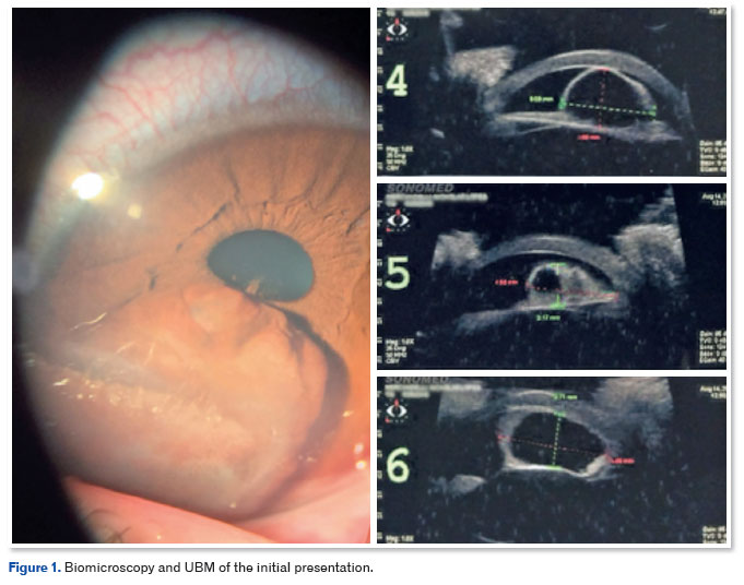
Initial treatment consisted of the application of Nd:YAG laser for anterior cystotomy, using low-energy parameters (single shots, with power progressively adjusted until rupture of the anterior cyst wall, without the need for a high secondary energy load). The procedure was performed under sedation on an outpatient basis, resulting in immediate reduction of the cystic content and opening of the peripheral anterior chamber angle, with no immediate pressure spikes or hyphema. In about 2 months, the patient presented with the first recurrence of the condition, with the cyst occluding about 90% of the pupillary axis. The patient then underwent surgical excision via dissection of the iris cyst with resection of the anterior leaf (anterior wall) and partial dissolution of posterior synechiae. The surgery was uneventful, resulting in an iris with a good appearance, a regular pupil, and, in the immediate postoperative period, only an inferior corneal leukoma (corneal scar/opacification) outside the visual axis.
Six months after the surgery, the patient presented with a new cyst recurrence, with corneal endothelial contact from the 6 to 8 o'clock position, associated with intermittent pain. Therefore, the patient underwent a new surgical reintervention involving a broad sector iridectomy from the 5 to 8 o'clock position, encompassing the lesion's base of implantation, via temporal, superior, and inferior corneal incisions, requiring a bimanual (two-port) anterior chamber aspiration to remove blood. The patient's postoperative course was marked by a well-appearing iris, with only a posterior synechia in the inferior region and no new cyst recurrences. Eighteen months after the last procedure, the patient presented with a localized anterior lens capsule opacity; best CVA was 20/25 in the right eye (OD) and 20/30 in the OS, and the condition was clinically followed up.
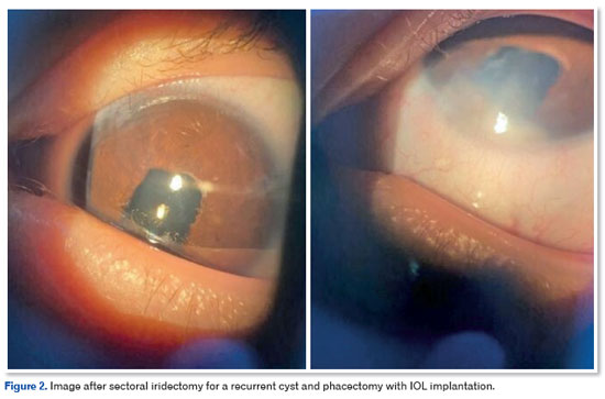
One year later, the patient presented with a 2/4+ cortical cataract compromising the visual axis and worsening of CVA in the OS to 20/100. A lensectomy was performed in the OS, requiring the use of iris hooks due to poor dilation, with implantation of an intraocular lens (IOL) in the capsular bag without intraoperative complications, followed 5 months later by an Nd:YAG laser posterior capsulotomy due to secondary capsular opacification. Seven months after the capsulotomy, at 6 years of age, the patient presented with a centrally clear cornea and no edema, a leucoma in the inferior portion, a formed anterior chamber, a well-appearing iris with an inferior iridectomy, a centered pupil, and a well-positioned IOL (Figure 2), with a CVA of 20/25 in the OD and 20/40 in the OS.
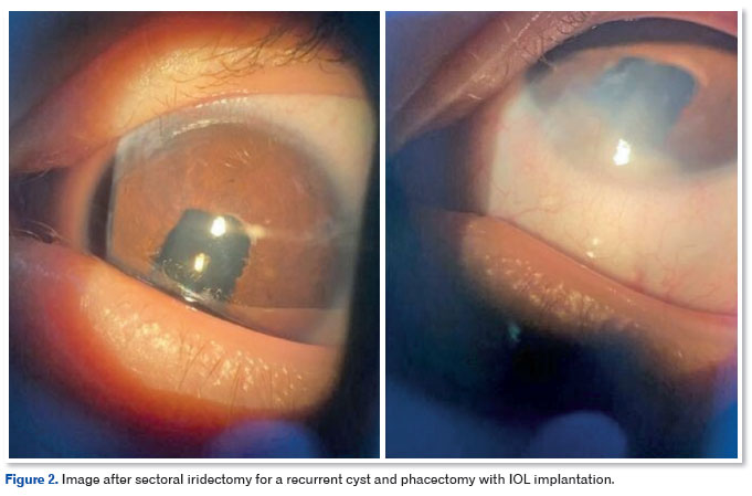
At the last examination, at 9 years of age, the patient maintained a CVA of 20/20 in the OD and 20/30 in the OS. Although the corneal endothelial cell density of the OS, assessed by specular microscopy, was significantly lower (1421 cells/mm2) compared to the contralateral eye (2926 cells/mm2), the cornea remained stable and without signs of edema or decompensation (Figure 3).
DISCUSSION
Historically, the differential diagnosis between cystic lesions and solid neoplasms of the anterior segment represented a significant diagnostic challenge2. Currently, the advent of high-resolution imaging technologies has transformed this approach. UBM has established itself as an indispensable tool, as its ability to emit high-frequency waves enables the delineation of retroiridian structures and the ciliary body, allowing for the precise measurement of the lesion's diameter and depth8. Concurrently, AS-OCT complements the diagnostic evaluation by providing micrometric details of the anterior surface and the cyst walls9. In the reported case, UBM was crucial not only for confirming the stromal nature of the pathology but also for mapping the lesion's close anatomical contact with the corneal endothelium and the lens, guiding subsequent surgical strategies.
The therapeutic management of primary iris stromal cysts is complex and varies according to the lesion's growth rate. While expectant management is supported in cases of indolent behavior and small dimensions — a pattern commonly observed in adults — the approach in children requires a lower threshold for intervention due to the speed at which ocular structures are affected in early childhood4. The indication for early surgery in these young patients is based on the need to preserve normal visual development and the anatomical integrity of the anterior segment1. In the present case, the natural course of the pathology confirmed this aggressive pattern, ratifying that a purely conservative approach would be insufficient.
Given the need for intervention, the initial choice of Nd:YAG laser is supported by previous studies, which describe the efficacy of this less invasive technique in adult patients with primary cysts (stromal or pigment epithelial), obtaining sustained regression of the cystic volume and visual improvement without complications10,11. However, in the present pediatric report, the benefit of laser cystotomy proved to be temporary, culminating in rapid recurrence within two months. This outcome resembled the findings of Shields et al.12, who, when evaluating a series of 16 patients with voluminous stromal cysts, observed failure in 100% of cases submitted to purely decompressive approaches, such as simple aspiration. The histopathological mechanism that justifies this early refractoriness lies in the fact that thermal opening (by laser) or needle aspiration are unable to eradicate the cyst's internal secretory epithelial cell lining, allowing the entrapped ectodermal cells to maintain active fluid production13.
Furthermore, the typical expansive behavior of stromal cysts in childhood — in contrast to their mostly stable nature in adults12 — explains why even the first surgical dissection with isolated resection of the anterior wall did not achieve the expected success, recurring after six months. This progression of refractoriness to laser and partial resection demonstrated that, to achieve resolution in complex pediatric cases, it becomes imperative to use techniques that permanently destroy or eliminate the secretory tissue. While Shields et al.12 advocate the combined use of sclerosing chemical agents (such as absolute alcohol) after the failure of simple aspiration, the present case was definitively resolved through a wide sector iridectomy, a surgical approach that proved definitive by removing en bloc the iris lesion's base of attachment.
The late development of secondary cataract in this patient can be postulated to result from a multifactorial mechanism correlated with the chronicity of the condition. The persistent contact exerted by the voluminous cyst promoted focal mechanical stress on the anterior capsule, altering its permeability and the metabolism of the local lenticular fibers14. Additionally, the history of multiple insults may have induced a low-grade tissue inflammatory response, accelerating opacification in childhood15. Therefore, the staged surgical management — progressing from laser to partial resection and, finally, to wide sectoral iridectomy with lensectomy/facectomy — proved effective in re-establishing the ocular anatomy and ensuring visual rehabilitation. Despite the substantial loss of endothelial cells in the OS, the maintenance of central corneal transparency and the final CVA of 20/30 at 9 years of age validate the positive impact of timely intervention and rigorous long-term follow-up in this complex pediatric case.
REFERENCES
1. Georgalas I, Petrou P, Papaconstantinou D, Brouzas D, Koutsandrea C, Kanakis M. Iris cysts: A comprehensive review on diagnosis and treatment. Surv Ophthalmol. 2018;63(3):347-364.
2. Hildreth T, Maino J, Hartong T. Primary and secondary iris cysts. J Am Optom Assoc. 1991;62(8):588-92.
3. Cronemberger S, Ferreira DM, Diniz Filho A, Mérula RV, Calixto N. Prevalência de cistos iridociliares em exames de biomicroscopia ultra-sônica. Arq Bras Oftalmol. 2006;69(4):471-475.
4. Shields JA, Shields CL, Lois N, Mercado G. Iris cysts in children: classification, incidence, and management. The 1998 Torrence A Makley Jr Lecture. Br J Ophthalmol. 1999;83(3):334-8.
5. Tavares PM, Herzog Neto G, Vilar EAG, Demori EM, Ventura MP, Solari HP. Iris cyst and atypical complications. Rev Bras Oftalmol. 2021;80(3):e0011.
6. Köse HC, Gündüz K, Hoşal MB. Iris Cysts: Clinical Features, Imaging Findings, and Treatment Results. Turk J Ophthalmol. 2020;50(1):31-36.
7. Ayres B, Allemann N, Nakanami C, Adan CBD. Cistos primários do epitélio pigmentar da íris e corpo ciliar: aspectos de biomicroscopia ultra-sônica. Arq Bras Oftalmol. 2000;63(5):337-43.
8. Augsburger JJ, Affel LL, Benarosh DA. Ultrasound biomicroscopy of cystic lesions of the iris and ciliary body. Trans Am Ophthalmol Soc. 1996;94:259-71.
9. Hau SC, Papastefanou V, Shah S, Sagoo MS, Restori M, Cohen V. Evaluation of iris and iridociliary body lesions with anterior segment optical coherence tomography versus ultrasound B-scan. Br J Ophthalmol. 2015;99(1):81-6.
10. Landim G, Léda R, Sobrinho V. Cisto de íris estromal primário em adulto tratado com Nd:YAG laser: relato de caso. eOftalmo. 2019;5(3):149-53.
11. Costa JCL, Montenegro ACP, Santos AV, Dias MBC, Dias TJ, Stenio R. Cistos irianos primários sintomáticos tratados com fotocoagulação a laser de Nd:YAG de dupla frequência. Rev Bras Oftalmol. 2017;76(4):198-201.
12. Shields CL, Arepalli S, Lally EB, Lally SE, Shields JA. Iris Stromal Cyst Management With Absolute Alcohol–Induced Sclerosis in 16 Patients. JAMA Ophthalmol. 2014;132(6):703-8.
13. Shields JA, Kline MW, Augsburger JJ. Primary iris cysts: a review of the literature and report of 62 cases. Br J Ophthalmol. 1984;68(3):152-66.
14. Mahjoub A, Abdesslem NB, Jouini A, Abderrazek AB, Ghorbel M, Mahjoub H. Surgical management of post-traumatic iris cyst: A case report. Int J Surg Case Rep. 2023 Apr:105:108037.
15. Budhwani M, Potti S. Traumatic cataract with iris cyst - A combined approach. Indian J Ophthalmol Case Rep. 2022;2(1):269-70.
| AUTHOR'S INFORMATION |
|
 |
»Gustavo Henrique M. R. Montalvão https://orcid.org/0000-0001-8310-0469 http://lattes.cnpq.br/4284733012830478 |
 |
» Manuella Neves da R. Nader https://orcid.org/0009-0002-3099-730X http://lattes.cnpq.br/7788292000906806 |
 |
» Cassiano Rodrigues Isaac https://orcid.org/0000-0002-4872-1451 http://lattes.cnpq.br/0982856065472123 |
 |
» Gilson de Santana Menezes Junior https://orcid.org/0009-0002-9359-0275 http://lattes.cnpq.br/7073052529955519 |
 |
» Vicente de Paula Freire da Silva Júnior https://orcid.org/0000-0002-2021-3818 http://lattes.cnpq.br/8490622827213279 |
 |
» João Lucas Carvalho Achkar https://orcid.org/0009-0007-8779-6142 http://lattes.cnpq.br/6330181327962834 |
 |
» Stella Cristina Alves Monreal https://orcid.org/0009-0006-3410-3552 http://lattes.cnpq.br/0419830954067675 |
Funding: None declared.
Conflicts of interest: None declared.
Received on:
March 26, 2026.
Accepted on:
July 14, 2026.
 eOftalmo está licenciada com uma Licença Creative Commons Atribuição-NãoComercial 4.0 Internacional.
 © 2026 Todos os Direitos Reservados


 Ler em português
 Português PDF
 Imprimir
 Enviar este artigo por email
 Como citar este artigo
 Enviar um comentário
 Mendeley
 Pocket
 Compartilhar no Facebook
 Compartilhar no Twitter
 Compartilhar no Linkedin
 Compartilhar no WhatsApp