

Fernanda Belga Ottoni Porto1,2,3; Rafael Mourão Agostini3,4,5; Raphael Stehling Fernandes5; Ana Paula Carneiro Rodrigues1; Julia Clara Dias Norberto Ferreira1; Fabrício Ribeiro Laender5; Rodrigo dos Anjos Versiani6; Aliança Profissional para Terapia Gênica
DOI: 10.17545/eOftalmo/2023.0042
Este artigo pertence à Edição Especial Dominando a arte da cirurgia vitreorretiniana: técnicas e dicas
Luxturna® (voretigene neparvovec-rzyl) is the first FDA- and Anvisa-approved gene therapy designed to correct RPE65 gene mutations associated with Leber congenital amaurosis (LCA) and retinitis pigmentosa. This groundbreaking treatment not only holds promise for restoring sight but also represents a paradigm shift in the way we approach inherited genetic disorders.
In this video, we present the first Luxturna® therapy performed by our group in Belo Horizonte, Minas Gerais, Brazil. The GeneTherapy Professional Alliance is the first gene therapy center in Minas Gerais and the third in South America. This is also the first Luxturna® treatment outside of the state of Sao Paulo in Brazil. We show the right eye of an 18-year-old woman who presented with vision loss since birth. At two years old, she was diagnosed by doctor Fernanda Porto with LCA. Biallelic pathogenic variants in the RPE65 were identified when the patient was 16 years old. The patient experienced progressive loss of central and peripheral vision as well as night vision. The retinas were translucent, the retinal vessels were attenuated, and the retina was relatively preserved at the posterior pole, but macular atrophy was still present inferiorly in both eyes affecting the parafovea, perifovea, and the foveal area. Photoreceptor cells were preserved in both eyes according to OCT. She also presented with keratoconus in both eyes.
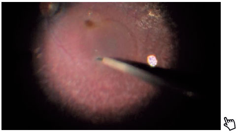
Initially, a standard four-port 23-gauge chandelier-assisted posterior vitrectomy was performed. We chose to stain the posterior hyaloid with triamcinolone acetonide to improve visualization because the posterior vitreous may be very adherent to the retina in young patients and therefore difficult to surgically detach. After core vitrectomy, we shaved the vitreous base under external scleral indentation. In this step, we also looked for peripheral lesions that could lead to retinal detachment. Luxturna® was prepared by the compound pharmaceutical team and delivered under sterile conditions to the operation room following industry guidelines. We used a microdose injection syringe with a 41-gauge subretinal cannula attached to the automated viscous fluid control. The retina was punctured using a 41-gauge cannula, and 0.3 ml of the medication were injected into the subretinal space, creating a bubble in the posterior pole. We also created a second bubble of 0.2 ml in the nasal retina to improve the temporal visual field of the patient. We performed a fluid-air exchange with a soft-tip cannula and closed all four scleral ports with Vycril 7-0 sutures.
Luxturna® therapy paves the way for an exciting future in the field of precision medicine and offers a ray of hope for those grappling with vision loss. Every gaze tells a story, and helping rewrite these narratives is heartening. May this milestone not only signify an achievement but, as technology continues to advance, also open the door for the development of similar therapies that target a broader spectrum of genetic disorders. We hope that questions on accessibility, affordability, and equitable distribution of such advanced therapies will be resolved in the near future, bringing hope to those who need it most.
REFERENCES
1. Russell S, Bennett J, Wellman J A, Chung D C, Yu Z F, Tillman A, et al. Efficacy and safety of voretigene neparvovec (AAV2-hRPE65v2) in patients with RPE65-mediated inherited retinal dystrophy: a randomized, controlled, open-label, phase 3 trial. Lancet. 2017;390(10097):849-860.
2. Davis J L, Gregori N Z, MacLaren R E, Lam B L. Surgical technique for subretinal gene therapy in humans with inherited retinal degeneration. Retina. 2019;39;Suppl 1:S2-S8.
3. Takahashi K, Morizane Y, Hisatomi T, Tachibana T, Kimura S, Hosokawa M M, et al. The influence of subretinal injection pressure on the microstructure of the monkey retina. PLOS ONE. 2018;13(12):e0209996.
4. Xue K, Groppe M, Salvetti A P, MacLaren R E. Technique of retinal gene therapy: delivery of viral vector into the subretinal space. Eye. 2017;31(9):1308-1316.
5. Scruggs B A, Huber Martins V Jr., Mariana Matioli da P, Katie K, Mark E P, Paul Y, et al. Injection pressure levels for creating blebs during subretinal gene therapy. Gene Ther. 2022 November; 29(10-11):601-607.
| AUTHORS INFORMATION |
|
 |
»Fernanda Belga Ottoni Porto https://orcid.org/0000-0002-4308-1766 http://lattes.cnpq.br/3705547122177092 |
 |
»Rafael Mourão Agostini http://lattes.cnpq.br/4837024324055533 https://orcid.org/0009-0004-8622-6218 |
 |
»Julia Clara Dias Norberto Ferreira http://lattes.cnpq.br/4624378177317170 https://orcid.org/0009-0002-5457-8847 |
 |
» Fabrício Ribeiro Laender http://lattes.cnpq.br/9393437510484424 https://orcid.org/0009-0001-8308-7572 |
 |
» Raphael Stehling Fernandes http://lattes.cnpq.br/2220253299512280 https://orcid.org/0009-0006-6117-2297 |
 |
» Ana Paula Carneiro Rodrigues http://lattes.cnpq.br/2529017243807277 https://orcid.org/0000-0002-9459-0835 |
 |
» Rodrigo dos Anjos Versiani http://lattes.cnpq.br/0589629829513519 https://orcid.org/0009-0001-2065-6491 |
Funding: No specific financial support was available for this study.
Conflict of interest: None of the authors have any potential conflict of interest to disclose.
Received on:
December 19, 2023.
Accepted on:
January 8, 2024.
 eOftalmo está licenciada com uma Licença Creative Commons Atribuição-NãoComercial 4.0 Internacional.
![]() © 2026 Todos os Direitos Reservados
© 2026 Todos os Direitos Reservados
 Ler em português
 Português PDF
 MP4
MP4
 Imprimir
 Enviar este artigo por email
 Como citar este artigo
 Enviar um comentário
 Mendeley
 Pocket