

Natália Fernandes Gonçalves; Luíza Marchesini Peixoto; Lais Soares de Carvalho; Rafaela Linck; Telma Rosana Virgens Gonzaga; Mário Henrique Camargos de Lima
DOI: 10.17545/eOftalmo/2021.0007
ABSTRACT
This study aimed to describe an unusual clinical case that was difficult to diagnose. We report the case of a child with a periorbital lesion who was being treated with oral antibiotics due to the initial diagnosis of periorbital cellulitis, with progressive clinical worsening. The identification of the epidemiological history and characteristics of the lesion were essential for correct diagnosis and effective treatment. Cutaneous sporotrichosis should thus be considered in the differential diagnosis of periorbital cellulitis, especially when associated with a history of previous contact with felines.
Keywords: Sporotrichosis; Sporothrix schenckii; Cutaneous; Ocular; Potassium iodide.
RESUMO
O objetivo deste estudo é descrever um caso clínico incomum e de difícil diagnóstico. Relatamos um caso de uma criança com lesão periorbitária que estava sendo tratada com antibióticos orais, devido ao diagnóstico inicial de celulite periorbitária bacteriana, com piora clínica progressiva. A história epidemiológica e a característica da lesão foram essenciais para o diagnóstico correto e para a instituição do tratamento eficaz. A esporotricose cutânea, desta forma, deve ser pensada como diagnóstico diferencial de celulite periorbitária, principalmente quando associado a história de contato prévio com felinos.
Palavras-chave: Sporotrichosis; Sporothrix schenckii; Cutaneus; Ocular; Potassium iodide.
INTRODUCTION
Sporotrichosis is a subacute or chronic fungal infection found worldwide but more frequently in temperate subtropical and tropical areas. It is considered as the most common subcutaneous mycosis in Latin America1.
Its main transmission route is the inoculation of the fungus into the skin through contact with soil, plants, or decaying materials, which is why it is known as “gardener’s disease”1,2. Facial involvement is more common in children due to the proximity between their faces and cats while playing with them3.
CASE REPORT
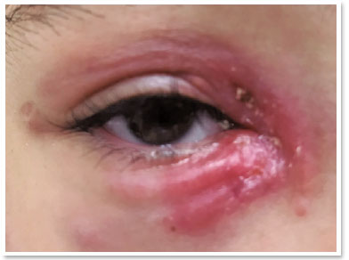
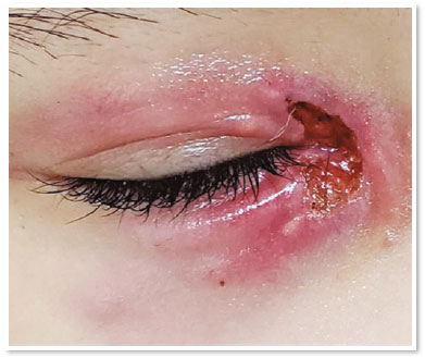
VAS, a 10-year-old female, was brought to the emergency room of the ophthalmology clinic at Complexo Hospitalar Padre Bento de Guarulhos (CHPBG) with complaints of hyperemia and periorbital edema in the right eye since six days. She had been using amoxicillin + clavulanate since a day. On examination, eyelid edema and hyperemia were observed. Visual acuity and ocular motility were preserved without any changes in biomicroscopy. The antibiotic use was maintained for seven days. The condition progressed to the development of pustules near the caruncle, with a slightly purulent discharge (Figure 1). The antibiotic was then replaced by oral cephalexin. The pustular lesions worsened, coalesced, and formed a deep purulent ulcerated lesion (Figure 2).
The patient was referred to the CHPBG dermatology team, who discovered recent injury to the right hand due to a scratch by her ill pet cat. Her pre-auricular lymph nodes were also palpable. We suspected a diagnosis of cutaneous sporotrichosis due to the lesion and epidemiological history. Initially, a skin biopsy was performed and oral potassium iodide was empirically prescribed. Biopsy results demonstrated suppurative granulomatous dermatitis and histochemical analysis did not show the presence of fungi and acid-alcohol fast bacilli (Ziehl-Neelsen, Grocott, PAS) on hematoxylin and eosin staining.
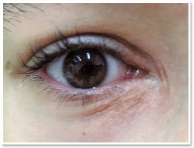
The lesion remitted completely after two months of treatment, with no reported side effects (Figure 3). The patient remained under treatment for another four months with gradual reduction of the potassium iodide dosage.
DISCUSSION
Sporotrichosis is a mycosis caused by different species of Sporothrix, classified into five classes: Sporothrix brasiliensis (Class I), Sporothrix schenckii (Class II), Sporothrix globosa (Class III), Sporothrix mexicana (Class IV), and Sporothrix pallida, formally known as S. albicans (Class V)1,2,4.
Infections peak during autumn and winter, due to increased relative humidity in endemic countries. Transmission occurs either by traumatic inoculation of contaminated organic materials or by animals, which act as vectors, such as rats, mice, and squirrels.
It is important to highlight that this condition often turns into an epidemic in Rio de Janeiro and parts of southern Brazil due to spread of infection by contaminated cats and affects a significant part of the population4,5. After inoculation, the incubation period in humans can range from a week to two months. It can be manifested in lymphocutaneous (most common), fixed or disseminated cutaneous, or extracutaneous forms. It begins as a small hardened papule, which progresses into a nodule and an ulcerated lesion3. The disseminated cutaneous form is rare, initially asymptomatic, more frequent among immunosuppressed individuals, of hematogenous dissemination, and may cause lung diseases, sinusitis, and meningitis6,7. Isolating Sporothrix in a culture from clinical specimens such as exudate, sputum, or lesion scraping is the gold standard for diagnosis. The “asteroid body” is a structure considered typical but not pathognomonic. Incubation in sheep blood agar can show the growth of colonies of “cigar-shaped bodies”, whereas Sabouraud dextrose agar can feature a “salt and pepper appearance,” highly suggestive of Sporothrix schenckii2.
Histology can guide the diagnosis, but is not pathognomonic. Generally, an inflammatory reaction is observed, with suppurative granulomatous nodules, which may be confused with tuberculosis, other mycoses, and syphilis. Organisms are scarce and rarely detected in histopathology2. Thus, as observed in our case, the clinical aspect of the lesions and the epidemiological link became essential for adequate diagnosis and treatment, as biopsy results are negative in many cases.
Serological (direct immunofluorescence and ELISA), molecular (PCR), and intradermal tests are alternatives used to diagnose the disease. The latter is highly useful in epidemiological surveys for studies on prevalence in certain geographical areas7. The treatment of choice is oral itraconazole for three to six months, but potassium iodide is also a low-cost and highly efficient option. However, compared with itraconazole, potassium iodide has more side effects, such as metallic taste, nausea, vomiting, anorexia, epigastralgia, and diarrhea3,4. Prolonged use may even cause potassium toxicity (arrhythmias, weakness, mental confusion, hand paresthesia) in some cases5,8.
This case report aimed to broaden the spectrum of differential diagnoses for periorbital cellulitis. Anamnesis and epidemiological history play a fundamental role in clinical diagnosis, especially in lesions refractory to initial treatment.
REFERENCES
1. Vieira-Dias D, Sena CM, Oréfice F, Tanure MA, Hamdan JS. Ocular and concomitant cutaneous sporotrichosis. Mycoses. 1997; 40(5-6):197-201.
2. Ochoa-Reyes J, Ramos-Martínez E, Treviño-Rangel R, González GM, Bonifaz A. Esporotricosis del pabellón auricular. Comunicación de un caso atípico simulando una celulitis bacteriana. Rev Chil Infectol. 2018;35(1):83-87.
3. Estrada-Castañón R, Chávez-López G, Estrada-Chávez G, Bonifaz A. Report of 73 cases of cutaneous sporotrichosis in Mexico. An Bras Dermatol. 2018;93(6):907-9.
4. Rees RK, Swartzberg JE. Feline-transmitted sporotrichosis: A case study from California. Dermatol Online J. 2011;17(6):2.
5. Mahlberg MJ, Patel R, Rosenman K, Cheung W, Wang N, Sanchez M. Fixed cutaneous sporotrichosis. Dermatol Online J. 2009;15(8):5.
6. García-Malinis AJ, Milagro Beamonte A, Torres Sopena L, García-Callen O, Puertolas-Villacampa P, Gilaberte Y. Cutaneous sporotrichosis treated with methylene blue-daylight photodynamic therapy. J Eur Acad Dermatol Venereol. 2018; 32(3):e90-e91.
7. Medeiros KB, Landeiro LG, Diniz LM, Falqueto A. Disseminated cutaneous sporotrichosis associated with ocular lesion in an immunocompetent patient. An Bras Dermatol. 2016;91(4):537-9.
8. Bonifaz A, Tirado-Sánchez A. Cutaneous Disseminated and Extracutaneous Sporotrichosis: Current Status of a Complex Disease. J Fungi (Basel). 2017;3(1):6.
AUTHOR’S INFORMATION


Funding: No specific financial support was available for this study
Disclosure of potential conflicts of interest: None of the authors have any potential conflict of interest to disclose
Received on:
October 13, 2020.
Accepted on:
September 26, 2020.
 eOftalmo está licenciada com uma Licença Creative Commons Atribuição-NãoComercial 4.0 Internacional.
![]() © 2026 Todos os Direitos Reservados
© 2026 Todos os Direitos Reservados

 Ler em português
 Português PDF
 Imprimir
 Enviar este artigo por email
 Como citar este artigo
 Enviar um comentário
 Mendeley
 Pocket