

Guilherme Gouveia Hollunder; Arthur Moreira de Freitas; Amanda Simonic; Tarcísio Veloso; Leonardo Coelho Gontijo
DOI: 10.17545/eOftalmo/2026.v12.0013
ABSTRACT
PURPOSE: This report describes a case of ocular sporotrichosis and emphasizes its clinical and epidemiological importance in the context of the rising incidence of zoonotic transmission in Brazil.
METHODS: We present the clinical course, diagnostic evaluation, and treatment of a 30-year-old woman diagnosed with granulomatous conjunctivitis caused by Sporothrix schenckii (S. schenckii) infection.
RESULTS: The patient presented with a granulomatous lesion in the inferior conjunctival fornix of the left eye, accompanied by conjunctival hyperemia and localized discomfort. Microbiological examination of conjunctival scraping confirmed the presence of S. schenckii. Treatment with oral itraconazole (200mg/day) was initiated, resulting in progressive lesion regression within 1 month. Complete clinical resolution was achieved after 90 days, with no recurrence during follow-up and preservation of visual acuity.
CONCLUSION: Although uncommon, ocular sporotrichosis is increasingly recognized in hyperendemic regions of Brazil due to zoonotic transmission. Early clinical suspicion in cases of granulomatous conjunctivitis and prompt antifungal therapy are essential for favorable outcomes.
Keywords: Conjunctivitis; Sporotrichosis; Ocular fungal infections; Sporothrix schenckii; Zoonoses.
RESUMO
OBJETIVO: Relatar um caso raro de esporotricose ocular e destacar sua relevância clínica e epidemiológica no contexto do aumento da incidência de transmissão zoonótica no Brasil.
MÉTODOS: Descrevemos a apresentação clínica, a propedêutica e o manejo terapêutico de uma mulher de 30 anos com conjuntivite granulomatosa por Sporothrix schenckii.
RESULTADOS: A paciente apresentou lesão granulomatosa no fórnice inferior do olho esquerdo, associada a hiperemia conjuntival e desconforto local. A cultura do raspado conjuntival confirmou S. schenckii. Foi iniciado Itraconazol oral 200mg/dia, com regressão gradual da lesão após um mês. A resolução completa, com cicatrização, ocorreu em 90 dias, sem recorrência durante o seguimento e com preservação da acuidade visual.
CONCLUSÃO: A esporotricose ocular, embora incomum, tem emergido em regiões brasileiras hiperendêmicas devido à transmissão zoonótica. A suspeição diagnóstica frente a conjuntivites granulomatosas e o tratamento adequado são fundamentais.
Palavras-chave: Conjuntivite; Esporotricose; Infecções oculares fúngicas; Sporothrix schenckii; Zoonoses.
INTRODUCTION
Human sporotrichosis is a subcutaneous mycosis caused by fungi of the genus Sporothrix, most commonly S. schenckii, and is recognized as the most prevalent subcutaneous fungal infection worldwide1. The disease occurs more frequently in tropical and subtropical regions, including Latin America, Asia, and Africa2.
Traditionally, infection occurs following traumatic inoculation of the skin through contact with plant material, organic debris, or contaminated soil. However, since the early 2000s, an increasing number of cases have been linked to animal scratches and bites, particularly from cats, indicating a shift toward zoonotic transmission and contributing to outbreaks in endemic regions3.
Clinically, sporotrichosis most commonly manifests as cutaneous disease, typically in the form of ascending nodular lymphangitis. This presentation is characterized by subcutaneous nodular lesions that may become ulcerated and form draining sinuses with purulent discharge4–6. Nevertheless, extracutaneous manifestations may also occur, including osteoarticular, pulmonary, meningeal, and ocular involvement4–6.
Although ocular involvement is uncommon, cases of ocular sporotrichosis have been increasingly reported in hyperendemic regions, particularly in association with the rising incidence of zoonotic transmission2,3.
This report describes a rare case of granulomatous conjunctivitis caused by S. schenckii. managed in a tertiary hospital in Belo Horizonte, Brazil, highlighting its diagnostic approach and clinical management in the context of the increasing burden of sporotrichosis in the country.
CASE REPORT
A 30-year-old woman was referred to the ophthalmology emergency service due to a granulomatous lesion in the inferior conjunctival fornix of the left eye. The patient reported that the lesion had appeared 45 days prior and had progressively worsened, without significant pain, being associated only with mild pruritus. She denied systemic symptoms, comorbidities, and any prior ophthalmological history. However, she reported a history of close contact with cats. On examination, best-corrected visual acuity (pinhole) was 20/20 in both eyes. Slit-lamp examination of the right eye was unremarkable. The left eye showed mild conjunctival hyperemia, a clear cornea, and a hyperemic, nodular lesion measuring approximately 11 × 2 mm with a whitish central area, located in the inferior conjunctival fornix (Figure 1).
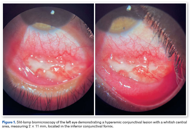
Given the clinical suspicion of ocular sporotrichosis, a conjunctival scraping culture was obtained. Antifungal therapy was deferred pending microbiological confirmation. After 1 month, the culture yielded fungal growth identified as S. schenckii. Treatment with oral itraconazole (200mg/day) was initiated for 90 days, with monthly monitoring of hepatic function. A marked reduction in lesion size was observed after 1 month of therapy, and complete clinical resolution was achieved at the end of treatment (Figure 2).
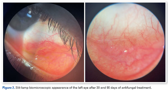
DISCUSSION
Clinically, granulomatous conjunctivitis associated with sporotrichosis typically presents as nodular or tumoral conjunctival lesions, often salmon-colored and of granulomatous appearance. These lesions may be accompanied by conjunctival hyperemia and edema, and in some cases, ipsilateral preauricular lymphadenopathy, consistent with Parinaud oculoglandular syndrome4–6. Diagnosis is often challenging because the clinical presentation can mimic other causes of granulomatous conjunctivitis, including tuberculosis, sarcoidosis, and bacterial or viral infections. Histopathological examination typically demonstrates granulomatous inflammation, sometimes with microabscess formation and spherical yeast-like structures, while definitive diagnosis is established through fungal culture or molecular detection. Granulomatous conjunctivitis from conjunctival specimens1,4-6.
Ocular sporotrichosis may occur in both immunocompetent and immunocompromised individuals. The use of topical or systemic corticosteroids has been implicated as a potential predisposing factor or may exacerbate disease progression4. First-line treatment is oral itraconazole, which may be combined with topical or subconjunctival antifungal therapy in severe or refractory cases. Clinical response is generally favorable, with lesion resolution occurring over weeks to months following appropriate therapy1,4-6.
Although uncommon, ocular sporotrichosis has been increasingly reported in hyperendemic regions, paralleling the rise in zoonotic transmission of the disease³.
In recent years, Brazil has reported an increase in cases of sporotrichosis in various regions of the country2–3. A study conducted in the Rio de Janeiro Metropolitan Region analyzed 9,552 suspected human cases and 12,532 animal cases, evidencing spatial expansion and urban hyperendemicity between 2013 and 20207. Another study conducted in the state of Amazonas reported 950 human cases and 2,823 animal cases, with a 304% increase in human cases from 2021 to 2022 and a 249% increase from 2022 to 20238. In the state of Paraná, located in the southern region of the country, where the disease has been included in the list of compulsory notification since 2020, the number of human cases increased from 253 in 2022 to 853 in 2023; among felines, reported cases rose from 1,412 in 2022 to 3,290 in 20239.
In Brazil, mandatory reporting of sporotrichosis is not yet implemented at the national level, being required only in some states and municipalities. This fact contributes to underreporting, hinders real-time understanding of the epidemiological situation, and delays the implementation of control measures, which favors the maintenance of prevalence and incidence in certain regions2-3.
This case illustrates the importance of maintaining diagnostic suspicion in the presence of granulomatous conjunctivitis in endemic areas. Furthermore, it is necessary to evaluate the inclusion of sporotrichosis in the list of nationally notifiable diseases to enable better management in areas of epidemiological risk.
AUTHOR CONTRIBUTIONS:
Significant contribution to the conception and design of the study: Guilherme Gouveia Hollunder; Arthur Moreira de Freitas.
Data acquisition: Guilherme Gouveia Hollunder; Arthur Moreira de Freitas; Amanda Simonic; Tarcísio Veloso.
Data analysis and interpretation: Guilherme Gouveia Hollunder; Arthur Moreira de Freitas; Amanda Simonic; Tarcísio Veloso.
Manuscript drafting: Guilherme Gouveia Hollunder; Arthur Moreira de Freitas.
Critical revision of important intellectual content: Leonardo Coelho Gontijo.
Final approval of the version to be submitted: Guilherme Gouveia Hollunder; Arthur Moreira de Freitas; Amanda Simonic; Tarcísio Veloso; Leonardo Coelho Gontijo.
Statistical analysis: Guilherme Gouveia Hollunder; Arthur Moreira de Freitas; Amanda Simonic; Tarcísio Veloso.
Funding: Not applicable.
Supervision of administrative, technical, or material support: Leonardo Coelho Gontijo.
Research group leadership: Leonardo Coelho Gontijo.
REFERENCES
1. Silva NF, Gadelha C, Silva L, Pazos N, Silva A, Silva M, et al. Case report: ocular sporotrichosis – a seven-case series. Am J Trop Med Hyg. 2023;108(6):1109-1114.
2. Alvarez CM, Oliveira MME, Pires RH. Sporotrichosis: a review of a neglected disease in the last 50 years in Brazil. Microorganisms. 2022;10(11):2152.
3. Freire CECA, Reis RM. Reemergence of zoonotic sporotrichosis in Brazil as a public health threat. Discover Public Health. 2025;22:187.
4. Su Q, Dong H, Cui L, Zhang L. Atypical conjunctival sporotrichosis secondary to Mooren's ulcer: a case report. BMC Infect Dis. 2025;25(1):71.
5. Hampton DE, Adesina A, Chodosh J. Conjunctival sporotrichosis in the absence of antecedent trauma. Cornea. 2002;21(8):831-3.
6. Kashima T, Honma R, Kishi S, Hirato J. Bulbar conjunctival sporotrichosis presenting as a salmon-pink tumor. Cornea. 2010; 29(5):573-6.
7. Ferreira VCD, Figueiredo ABF, Magalhães MAFM, Pereira AS, Tassinari W. Temporal and spatial distribution of sporotrichosis in the Rio de Janeiro Metropolitan Area, Brazil: a comparison between human and animal cases (2013–2020). Cad Saúde Pública. 2025;41(2):e00133024.
8. Mesquita VA, Talhari S, Leturiondo AL, Souza GC, Brito EM, Andrade SL, et al. Zoonotic Sporotrichosis outbreak: Emerging public health threat in the Amazon State, Brazil. PLoS Negl Trop Dis. 2024;18(7):e0012328.
9. Secretaria de Estado da Saúde do Paraná. Esporotricose no Paraná: situação epidemiológica e medidas de controle. Curitiba: SESA-PR; 2024. Available from: https://www.saude.pr.gov.br/Pagina/Esporotricose
| AUTHOR INFORMATION |
|
 |
» Guilherme Gouveia Hollunder orcid.org/0000-0002-4948-2472 lattes.cnpq.br/7357322782551551 |
 |
» Arthur Moreira de Freitas orcid.org/0009-0002-2740-3865 lattes.cnpq.br/2721707151901421 |
 |
» Amanda Simonic orcid.org/0009-0001-4920-8377 lattes.cnpq.br/7357322782552223 |
 |
» Tarcísio Veloso Rabelo orcid.org/0009-0009-5203-7319 lattes.cnpq.br/0911799108595422 |
 |
» Leonardo Coelho Gontijo orcid.org/0009-0000-8520-5010 lattes.cnpq.br/3690478000038886 |
Funding: No specific financial support was available for this study.
Approved by the following Research Ethics Committee: Dr. Francisco das Chagas Lima e Silva – Santa Casa de Misericórdia de Belo Horizonte – SCMBH (CAAE: 865).
Data Availability Statement: The data generated and/or analyzed during t
Conflict of interest: None of the authors have any potential conflict of interest to disclose.
Received on:
October 14, 2025.
Accepted on:
June 15, 2026.
 eOftalmo está licenciada com uma Licença Creative Commons Atribuição-NãoComercial 4.0 Internacional.
 © 2026 Todos os Direitos Reservados


 Ler em português
 Português PDF
 Imprimir
 Enviar este artigo por email
 Como citar este artigo
 Enviar um comentário
 Mendeley
 Pocket
 Compartilhar no Facebook
 Compartilhar no Twitter
 Compartilhar no Linkedin
 Compartilhar no WhatsApp