

Luiza Carneiro Bertazzi1; Júlia Margoni Biluca1; Daniel Haruo Ishigai1; Patrícia Lury Fuke Yoshiyasu1; Ricardo Tomoyoshi Kanecadan1; Renan Carneiro Nogueira Bertazzi2
DOI: 10.17545/eOftalmo/2026.v12.0012
ABSTRACT
PURPOSE: This study aimed to evaluate the surgical outcomes of external dacryocystorhinostomy performed at a teaching hospital over a two-year period and to identify factors associated with surgical success.
METHODS: This retrospective observational analytical study reviewed the medical records of patients aged ≥18 years who underwent external dacryocystorhinostomy at the Department of Ophthalmology, Santa Casa de Misericórdia de São Paulo, Brazil, between July 2020 and July 2022. Demographic characteristics, clinical findings, surgical variables, and postoperative outcomes were assessed. Surgical success was defined as either resolution of preoperative symptoms or normalization of the fluorescein disappearance test at the 6-month postoperative follow-up, as documented in the medical records. Statistical analyses were performed using the chi-square test, Student's t-test, and Fisher's exact test, with statistical significance set at p<0.05.
RESULTS: A total of 102 external DCR procedures met the inclusion criteria and were included in the analysis. Surgical success was achieved in 91 cases (89.2%), whereas surgical failure occurred in 11 cases (10.8%). Among the variables evaluated, only the use of a lacrimal stent was significantly associated with a higher rate of surgical success (p<0.001). No significant associations were identified between surgical outcomes and patient age, sex, laterality, reflux on lacrimal sac compression, previous episodes of acute dacryocystitis, or the presence of comorbidities.
CONCLUSIONS: External dacryocystorhinostomy demonstrated a high success rate when performed in a teaching hospital setting. The use of a lacrimal stent was the only factor significantly associated with improved surgical outcomes. These findings support the effectiveness and reproducibility of external dacryocystorhinostomy, even when performed by surgeons in training under appropriate supervision.
Keywords: Dacryocystorhinostomy; Nasolacrimal duct obstruction; Lacrimal apparatus diseases; Hospitals, teaching.
RESUMO
OBJETIVO: Avaliar os desfechos cirúrgicos da dacriocistorrinostomia externa (DCR) realizada em um hospital de ensino ao longo de um período de dois anos e identificar fatores associados ao sucesso cirúrgico.
MÉTODOS: Foi realizado um estudo retrospectivo, observacional e analítico, baseado na análise de prontuários médicos de pacientes com idade ≥ a 18 anos submetidos à dacriocistorrinostomia externa no Departamento de Oftalmologia da Santa Casa de Misericórdia de São Paulo, Brasil, entre julho de 2020 e julho de 2022. Foram analisados dados demográficos, características clínicas, variáveis cirúrgicas e desfechos pós-operatórios. O sucesso cirúrgico foi definido pela resolução dos sintomas pré-operatórios ou pela normalização do teste de desaparecimento da fluoresceína no seguimento pós-operatório de seis meses, conforme registro em prontuário. As análises estatísticas foram realizadas utilizando os testes do qui-quadrado, t de Student e exato de Fisher, adotando-se nível de significância de p<0,05.
RESULTADOS: Um total de 102 procedimentos de DCR externa preencheram os critérios de inclusão e foram incluídos na análise. O sucesso cirúrgico foi alcançado em 91 casos (89,2%), enquanto 11 pacientes (10,8%) apresentaram falha cirúrgica. Entre as variáveis avaliadas, apenas o uso de stent lacrimal esteve significativamente associado a maiores taxas de sucesso cirúrgico (p<0,001). Não foram observadas associações estatisticamente significativas entre os desfechos cirúrgicos e idade, sexo, lateralidade, presença de refluxo à compressão do saco lacrimal, episódios prévios de dacriocistite aguda ou comorbidades.
CONCLUSÕES: A dacriocistorrinostomia externa demonstrou alta taxa de sucesso quando realizada em ambiente de hospital de ensino. O uso de stent lacrimal foi o único fator significativamente associado à melhora dos desfechos cirúrgicos. Esses achados reforçam a eficácia e a reprodutibilidade da DCR externa, mesmo quando realizada por cirurgiões em treinamento sob supervisão adequada.
Palavras-chave: Dacriocistorrinostomia; Obstrução do ducto lacrimonasal; Doenças do aparelho lacrimal; Hospitais de ensino.
INTRODUCTION
Dacryocystorhinostomy (DCR) was first described by Toti in 1904 as a surgical procedure designed to establish a direct communication between the lacrimal sac and the nasal cavity, thereby restoring lacrimal drainage1. The technique was subsequently incorporated into the classical literature on lacrimal drainage disorders and became the standard surgical treatment for distal lacrimal system obstruction2. With advances in endonasal surgery, alternative approaches for managing lacrimonasal obstructions have emerged, including endoscopic DCR, which has demonstrated favorable long-term outcomes3. Nevertheless, endoscopic DCR requires a specific learning curve and depends heavily on the surgeon's familiarity with nasal anatomy, endoscopic equipment, and the technical aspects of the procedure4.
Despite the growing use of endoscopic techniques, external DCR remains a widely accepted treatment for acquired nasolacrimal duct obstruction in adults because of its consistently high functional success rates and satisfactory cosmetic outcomes. Previous studies evaluating skin incisions in external DCR have shown that carefully planned surgical approaches can produce minimally visible scars while maintaining excellent functional results and high levels of patient satisfaction5,6. Assessment of surgical outcomes in teaching hospitals is particularly important because it provides insight into the effectiveness and reproducibility of the procedure within a supervised training environment7. Furthermore, Brazilian studies have highlighted the clinical relevance of dacryostenosis and nasolacrimal duct obstruction in routine ophthalmic practice8.
In addition to technical considerations related to the skin incision, osteotomy, and construction of mucosal flaps, the role of silicone lacrimal intubation in external DCR remains controversial. Systematic reviews and comparative studies suggest that the effect of silicone stenting may vary according to surgical technique, patient characteristics, and study methodology. However, some evidence indicates a potential benefit in selected cases undergoing external DCR9. Reports from university-based centers have demonstrated that external DCR can achieve success rates comparable to those reported by other specialized institutions10. Likewise, studies evaluating endoscopic DCR have shown that factors such as patient age, surgical technique, and institutional experience may influence postoperative outcomes11,12. Analyses of the learning curve associated with lacrimal procedures further emphasize the importance of critically assessing surgical performance in training centers13.
External DCR continues to hold particular clinical relevance because it provides direct visualization of the lacrimal sac, facilitates the creation of a sufficiently large osteotomy, and allows precise anatomical management of the mucosal flaps, all of which contribute to favorable surgical outcomes reported in the literature. Moreover, when the skin incision is appropriately selected and meticulously performed, the resulting scar is generally inconspicuous and should not, by itself, preclude the use of the external approach. In teaching hospitals, however, surgical outcomes may be influenced by both patient-related factors and the learning curve of surgeons in training, despite the presence of direct supervision throughout the procedure.
Therefore, the primary objective of this study was to evaluate the surgical outcomes of external dacryocystorhinostomy performed at a teaching hospital over a two-year period. The secondary objectives were to characterize the clinical and epidemiological profiles of patients undergoing the procedure, describe their principal clinical manifestations, and identify factors potentially associated with surgical success or failure.
METHODS
This retrospective observational analytical study evaluated patients treated at the Department of Ophthalmology, Santa Casa de Misericórdia de São Paulo, São Paulo, Brazil, who underwent external DCR between July 2020 and July 2022.
The study protocol was approved by the Research Ethics Committee of Santa Casa de Misericórdia de São Paulo (CEP No. 8.078.964; CAAE No. 84088923.9.0000.5479). Given the retrospective nature of the study, the requirement for informed consent was waived. All procedures were conducted in accordance with the ethical principles outlined in the Declaration of Helsinki. Data were obtained through a review of patients' medical records.
Eligible participants were adults aged ≥18 years of either sex with a diagnosis of lower nasolacrimal duct obstruction who underwent external DCR during the study period. Patients younger than 18 years and those presenting with upper (canalicular) obstruction, lacrimal sac tumors, facial anatomical abnormalities, or incomplete medical records were excluded from the analysis.
During the study period, 136 external DCR procedures were performed by the surgical team. After application of the inclusion and exclusion criteria, 102 medical records were considered eligible and were included in the final analysis.
Demographic characteristics, clinical history, findings from the lacrimal evaluation, and surgery-related variables were assessed. Clinical symptoms documented at the initial presentation included epiphora, ocular discharge, and edema in the lacrimal sac region. A history of acute dacryocystitis, systemic comorbidities, and the laterality of the obstruction were also evaluated. Lacrimal assessment findings included the presence of reflux upon compression of the lacrimal sac and the results of the fluorescein dye disappearance test. Among the surgical variables analyzed, the use of silicone lacrimal intubation was recorded.
For variables with unavailable information in the medical records, the data were categorized as "not reported." This category was retained in the tables to preserve the total number of cases analyzed and to explicitly indicate the presence of missing data, without imputation or redistribution of values across the remaining categories.
The fluorescein dye disappearance test (Milder's test) was performed during the preoperative assessment as an adjunctive tool for the evaluation of lacrimal drainage obstruction. The procedure consisted of instilling one drop of 1% fluorescein into the inferior conjunctival fornix, followed by assessment of dye retention within the tear meniscus after approximately 5 minutes. The test was considered positive when evident fluorescein retention was observed, indicating impaired lacrimal drainage, and negative when adequate clearance of the dye occurred.
Surgical outcomes were assessed at the 6-month postoperative follow-up visit. Surgical success was defined as the resolution of preoperative symptoms or normalization of the fluorescein dye disappearance test, as documented in the medical records. Persistent clinically significant epiphora, ocular discharge, edema in the lacrimal sac region, or fluorescein retention on functional testing was considered indicative of surgical failure.
During the 2-year study period, the surgeries were performed by first- and second-year fellows in the Oculoplastic, Orbital, and Lacrimal Fellowship Program under the supervision of preceptors and attending physicians of the service.
External DCR was performed under general anesthesia. Regarding the surgical technique, the procedure began with an approximately 10–15 mm skin incision made inferior to the medial canthal tendon, along the anterior lacrimal crest. After layer-by-layer dissection and exposure of the anterior lacrimal crest, an osteotomy was created using appropriate surgical instruments. Following exposure of the lacrimal sac and nasal mucosa, an anterior flap was fashioned by creating and suturing the anterior lacrimal sac and nasal mucosal flaps. After creation and suturing of the anterior flaps of the lacrimal sac and nasal mucosa, bicanalicular intubation with Silastic® silicone tubing was performed using a Crawford probe whenever technically feasible.
Postoperatively, patients received a combination antibiotic–corticosteroid eye drop regimen for 7 days with gradual tapering. The use of preservative-free lubricating eye drops was prescribed at a frequency of six instillations per day. Patients were instructed to perform nasal hygiene using saline solution. When performed, lacrimal intubation with a Silastic® silicone tube was maintained for a variable period ranging from 45 to 60 days and was subsequently removed in the outpatient setting.
The data were collected by a single researcher and tabulated using Microsoft Excel for analysis. Statistical analyses were performed using the chi-square test with Yates' correction, Student's t-test, and Fisher's exact test, as appropriate. A p-value <0.05 was considered statistically significant.
RESULTS
During the study period, 136 external dacryocystorhinostomy procedures were performed by the Lacrimal Pathway Team of the Department of Ophthalmology at Santa Casa de São Paulo. After application of the inclusion and exclusion criteria, 102 procedures were included in the analysis. Of the 34 excluded cases, nine had secondary causes of nasolacrimal obstruction, such as tumors or trauma, whereas the remaining cases were excluded because of incomplete clinical data or insufficient postoperative follow-up.
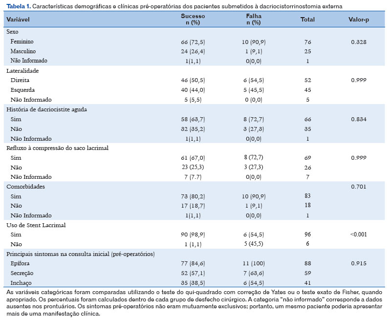
The clinical and demographic characteristics of the study population as well as the comparison between the surgical success and failure groups, are presented in T able 1. Female patients predominated, and unilateral involvement was more frequently observed. Epiphora was the most common clinical complaint at the initial evaluation.
After 6 months of follow-up, 91 procedures (89.2%) were classified as surgical successes, whereas 11 procedures (10.8%) were classified as surgical failures. Statistical analysis demonstrated that lacrimal intubation with a silicone stent was associated with a higher rate of surgical success (p<0.001). However, this finding should be interpreted with caution, as only six patients did not undergo stent placement because of intraoperative technical difficulties.
No significant differences were observed between the success and failure groups with respect to age, sex, laterality, reflux on compression of the lacrimal sac, previous episodes of acute dacryocystitis, associated comorbidities, or the primary clinical complaint (Table 1).
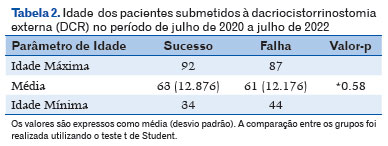
The age distribution of patients in the success and failure groups, including the minimum, mean, and maximum values, is presented in Table 2. No statistically significant difference in age was observed between the groups (p=0.58).
DISCUSSION
This study evaluated the outcomes of external dacryocystorhinostomy performed by surgeons in training under supervision in a teaching hospital setting. The surgical success rate was 89.2%, which is comparable to rates reported in previous external DCR series, where success rates generally range from approximately 85% to 95%. This finding suggests that, within a supervised environment, external DCR can achieve favorable surgical outcomes even when performed as part of a training program7,10-13.
External DCR remains a widely used surgical technique for the treatment of lower nasolacrimal duct obstruction. Its advantages include direct exposure of the lacrimal sac, the ability to create a large osteotomy, and the performance of the anastomosis under direct visualization. Furthermore, when the incision is carefully planned and positioned in the medial eyelid region or adjacent to the nasofacial sulcus, the resulting cutaneous scar tends to be inconspicuous and should not, in isolation, be considered a contraindication to the use of the external approach5,6.
The success rate observed in the present study was consistent with those reported in external DCR series conducted in different healthcare settings. This finding is particularly relevant because the procedures were performed in a teaching hospital by fellows under direct supervision. Thus, the results suggest that supervised surgical training can be associated with favorable outcomes, provided that appropriate technical standardization and adequate postoperative follow-up are maintained7,10.
The presence of reflux on compression of the lacrimal sac was analyzed because it is a frequent clinical finding in patients with nasolacrimal duct obstruction. This sign may indicate accumulation of secretions within the lacrimal sac and may reflect underlying anatomical alterations or chronic inflammatory changes of the lacrimal system. In the present study, however, reflux on compression was not significantly associated with either surgical success or failure.
The sample showed a predominance of female patients, with a mean age in the sixth decade of life, a profile consistent with that described in previous studies on nasolacrimal duct obstruction and DCR8,10,11. Nevertheless, neither sex nor age had a significant influence on surgical outcomes in this study.
The use of a lacrimal stent was associated with a higher surgical success rate in the statistical analysis performed. At the study institution, lacrimal intubation is routinely used during external DCR, reflecting the service protocol and surgical team experience. However, only six patients did not receive a stent due to intraoperative technical difficulties, rather than random allocation. Therefore, although a statistical association was observed between intubation and surgical success, the retrospective design does not allow a causal relationship between stent use and improved outcomes to be established. Previous studies have shown that the effect of intubation may vary depending on surgical technique, patient profile, and institutional experience9,10–13.
Among cases classified as surgical failures, six patients underwent revision surgery after the study period, whereas five opted for conservative management due to minimal or tolerable symptoms. This finding suggests that anatomical or functional failure does not necessarily correspond to clinically significant impairment for the patient, an aspect that should be considered when deciding on reintervention.
This study has limitations inherent to its retrospective design and reliance on medical record review, including dependence on the quality of clinical documentation and the absence of multivariate analysis. A statistical power calculation was not performed, and the small number of patients without a stent limited direct comparison between groups. Nevertheless, the observed results are consistent with those reported in the literature and suggest that external DCR may achieve a high success rate in a supervised surgical training setting.
In conclusion, this study demonstrated a surgical success rate of 89.2% for external dacryocystorhinostomy performed at a teaching hospital. These results are consistent with those reported in the literature and suggest that external DCR can yield favorable outcomes even in a supervised surgical training setting.
AUTHOR CONTRIBUTIONS:
Significant contributions to study conception and design: Luiza Carneiro Bertazzi and Patricia Lury.
Data collection: Julia Margoni Biluca. Analysis and interpretation of data: Ricardo Tomoyoshi Kanecadan. Manuscript preparation: Luiza Carneiro Bertazzi and Renan Carneiro Nogueira Bertazzi. Critical revision of the manuscript for important intellectual content: Daniel Haruo Ishigai. Final approval of the submitted manuscript: Luiza Carneiro Bertazzi, Ricardo Kanecadan, Patricia Lury, Julia Margoni Biluca, Daniel Haruo Ishigai, and Renan Carneiro Nogueira Bertazzi. Statistical analysis: Daniel Haruo Ishigai. Funding acquisition: Not applicable. Supervision of financial, technical, or logistical support: Julia Margoni Biluca. Study supervision and group leadership: Luiza Carneiro Bertazzi.
REFERENCES
1. Toti A. Nuovo metodo conservatore di cura radicale delle suppurazioni croniche del sacco lacrimale (dacriocistorinostomia). Clin Mod Firenze. 1904;10:385-389.
2. Duke-Elder S. Diseases of lacrimal passages: dacryocystitis. In: Duke-Elder S, editor. System of Ophthalmology: The Ocular Adnexa. Vol. 13. London: Henry Kimpton; 1974. p. 699-723.
3. Dietrich C, Mewes T, Kühnemund M, Hashemi B, Mann WJ, Amedee RG. Long-term follow-up of patients with microscopic endonasal dacryocystorhinostomy. Am J Rhinol. 2003;17(1):57-61.
4. Malhotra R, Norris JH, Sagili S, Al-Abbadi Z, Avisar I. The Learning Curve in Endoscopic Dacryocystorhinostomy: Outcomes in Surgery Performed by Trainee Oculoplastic Surgeons. Orbit. 2015;34(6):314-319.
5. Akaishi PMS, Mano JB, Pereira IC, Cruz AAV. Functional and cosmetic results of a lower eyelid crease approach for external dacryocystorhinostomy. Arq Bras Oftalmol. 2011;74(4):283-285.
6. Wadwekar B, Hansdak A, Nirmale SD, Ravichandran K. Cutaneous scar visibility after external dacryocystorhinostomy: A comparison of curvilinear and W shaped incision. Saudi J Ophthalmol. 2019;33(2):142-147.
7. Besharati MR, Rastegar A. Results and complications of external dacryocystorhinostomy surgery at a teaching hospital in Iran. Saudi Med J. 2005;26(12):1940-1944.
8. Schellini AS, Itoda LK, Moraes-Silva MRB. Dacryostenosis: analysis of cases treated in our service. Rev Bras Oftalmol. 1990;49:43-48.
9. Xie C, Zhang L, Liu Y, Ma H, Li S. Comparing the Success Rate of Dacryocystorhinostomy With and Without Silicone Intubation: A Trial Sequential Analysis of Randomized Control Trials. Sci Rep. 2017;7(1):1936.
10. Schellini SA, Sakamoto RH, Samahá JT, Arangon F, Padovani CR. Dacriocistorrinostomia externa em hospital universitário: avaliação dos resultados. Arq Bras Oftalmol. 2004;67(2):301-304.
11. Sung JY, Lee YH, Kim KN, Kang TS, Lee SB. Surgical outcomes of endoscopic dacryocystorhinostomy: analysis of age effect. Sci Rep. 2019;9(1):19861.
12. Saeed BM. Endoscopic DCR: a 10 years personal experience. Indian J Otolaryngol Head Neck Surg. 2019;71(Suppl 3):2039-2043.
13. Lee JJ, Lee HM, Lim HB, Seo SW, Ahn HB, Lee SB. Learning curve for endoscopic endonasal dacryocystorhinostomy. Korean J Ophthalmol. 2017;31(4):299-305.
| AUTHOR INFORMATION |
|
 |
» Luiza Carneiro Bertazzi https://orcid.org/0009-0002-1373-2780 http://lattes.cnpq.br/7468696785742243 |
 |
» Júlia Margoni Biluca https://orcid.org/0009-0004-3011-2641 http://lattes.cnpq.br/8564851077141443 |
 |
» Daniel Haruo Ishigai https://orcid.org/0000-0002-9192-8730 http://lattes.cnpq.br/1241273377602401 |
 |
» Patrícia Lury Fuke Yoshiyasu https://orcid.org/0009-0009-7336-4992 http://lattes.cnpq.br/9035314387729096 |
 |
» Ricardo Tomoyoshi Kanecadan https://orcid.org/0000-0003-3587-4229 http://lattes.cnpq.br/0215616887900199 |
 |
» Renan Carneiro Nogueira Bertazzi https://orcid.org/0009-0003-9606-1368 http://lattes.cnpq.br/3035187367135853 |
Funding: No specific financial support was available for this study.
Approved by the following Research Ethics Committee: Santa Casa de Misericórdia de São Paulo (CAAE: 84088923.9.0000.5479).
Data Availability Statement: The data generated and/or analyzed during the present study are included in the
Conflict of interest: None of the authors have any potential conflict of interest to disclose.
Received on:
February 25, 2026.
Accepted on:
June 15, 2026.
 eOftalmo está licenciada com uma Licença Creative Commons Atribuição-NãoComercial 4.0 Internacional.
 © 2026 Todos os Direitos Reservados


 Ler em português
 Português PDF
 Imprimir
 Enviar este artigo por email
 Como citar este artigo
 Enviar um comentário
 Mendeley
 Pocket
 Compartilhar no Facebook
 Compartilhar no Twitter
 Compartilhar no Linkedin
 Compartilhar no WhatsApp